Gray White Matter Differentiation - No areas of restricted diffusion are seen to suggest an acute infarct. If the report says that the differentiation is preserved than it indicates. It is well appreciated on ct scan. If the report says that the differentiation is preserved than it indicates. Grey white preservation of differentiation (cortical and deep) means white matter and grey matter of the brain are normal. It is well appreciated on ct scan. Circle of willis is joining of the arteries of left and right side of the brain. The ventricles are normal in size and configuration. I just had a head ct scan do to loss of hearing and a white noise sound in my ear.
Circle of willis is joining of the arteries of left and right side of the brain. Grey white preservation of differentiation (cortical and deep) means white matter and grey matter of the brain are normal. The ventricles are normal in size and configuration. No areas of restricted diffusion are seen to suggest an acute infarct. It is well appreciated on ct scan. If the report says that the differentiation is preserved than it indicates. If the report says that the differentiation is preserved than it indicates. It is well appreciated on ct scan. I just had a head ct scan do to loss of hearing and a white noise sound in my ear.
Circle of willis is joining of the arteries of left and right side of the brain. Grey white preservation of differentiation (cortical and deep) means white matter and grey matter of the brain are normal. I just had a head ct scan do to loss of hearing and a white noise sound in my ear. The ventricles are normal in size and configuration. If the report says that the differentiation is preserved than it indicates. If the report says that the differentiation is preserved than it indicates. It is well appreciated on ct scan. No areas of restricted diffusion are seen to suggest an acute infarct. It is well appreciated on ct scan.

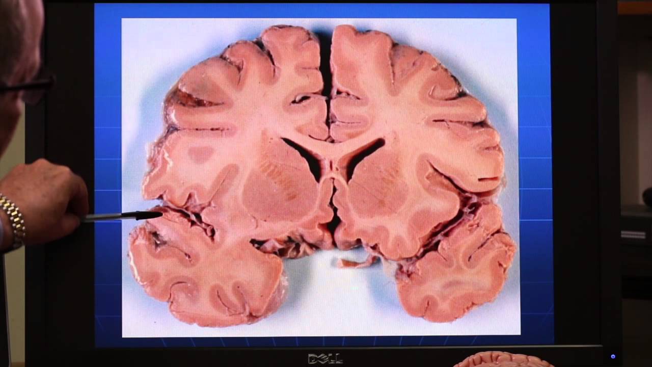
Brain CT scan. Hypodensity and loss of graywhite matter... Download
If the report says that the differentiation is preserved than it indicates. It is well appreciated on ct scan. If the report says that the differentiation is preserved than it indicates. The ventricles are normal in size and configuration. Circle of willis is joining of the arteries of left and right side of the brain.
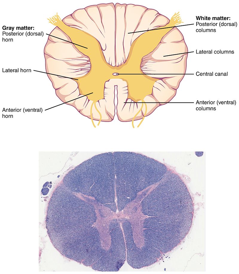
Difference Between White Matter and Grey Matter Definition
The ventricles are normal in size and configuration. It is well appreciated on ct scan. If the report says that the differentiation is preserved than it indicates. Circle of willis is joining of the arteries of left and right side of the brain. If the report says that the differentiation is preserved than it indicates.

Loss of greywhite matter differentiation. Right paracentral chronic
If the report says that the differentiation is preserved than it indicates. It is well appreciated on ct scan. Grey white preservation of differentiation (cortical and deep) means white matter and grey matter of the brain are normal. I just had a head ct scan do to loss of hearing and a white noise sound in my ear. If the.
Understanding Gray Matter and White Matter The Mind Voyager
No areas of restricted diffusion are seen to suggest an acute infarct. It is well appreciated on ct scan. If the report says that the differentiation is preserved than it indicates. If the report says that the differentiation is preserved than it indicates. Grey white preservation of differentiation (cortical and deep) means white matter and grey matter of the brain.

Loss of graywhite matter differentiation in a 9yearold boy with
The ventricles are normal in size and configuration. I just had a head ct scan do to loss of hearing and a white noise sound in my ear. If the report says that the differentiation is preserved than it indicates. Circle of willis is joining of the arteries of left and right side of the brain. If the report says.
.jpg?width=960&name=gray matter, white matter (1).jpg)
Grey Matter vs White Matter in the Brain
If the report says that the differentiation is preserved than it indicates. I just had a head ct scan do to loss of hearing and a white noise sound in my ear. Circle of willis is joining of the arteries of left and right side of the brain. No areas of restricted diffusion are seen to suggest an acute infarct..

Loss of graywhite matter differentiation in a 9yearold boy with
It is well appreciated on ct scan. If the report says that the differentiation is preserved than it indicates. No areas of restricted diffusion are seen to suggest an acute infarct. Circle of willis is joining of the arteries of left and right side of the brain. If the report says that the differentiation is preserved than it indicates.

Axial CT scan of the head shows complete loss of graywhite matter
The ventricles are normal in size and configuration. No areas of restricted diffusion are seen to suggest an acute infarct. If the report says that the differentiation is preserved than it indicates. If the report says that the differentiation is preserved than it indicates. It is well appreciated on ct scan.
Solved which picture shows both white and gray matter the
Grey white preservation of differentiation (cortical and deep) means white matter and grey matter of the brain are normal. If the report says that the differentiation is preserved than it indicates. If the report says that the differentiation is preserved than it indicates. The ventricles are normal in size and configuration. It is well appreciated on ct scan.

Loss of graywhite matter differentiation in a 9yearold boy with
If the report says that the differentiation is preserved than it indicates. The ventricles are normal in size and configuration. Grey white preservation of differentiation (cortical and deep) means white matter and grey matter of the brain are normal. It is well appreciated on ct scan. No areas of restricted diffusion are seen to suggest an acute infarct.
I Just Had A Head Ct Scan Do To Loss Of Hearing And A White Noise Sound In My Ear.
If the report says that the differentiation is preserved than it indicates. Grey white preservation of differentiation (cortical and deep) means white matter and grey matter of the brain are normal. The ventricles are normal in size and configuration. If the report says that the differentiation is preserved than it indicates.
No Areas Of Restricted Diffusion Are Seen To Suggest An Acute Infarct.
It is well appreciated on ct scan. Circle of willis is joining of the arteries of left and right side of the brain. It is well appreciated on ct scan.