Grey White Differentiation - Loss of this differentiation suggests the presence of oedema which may develop secondary to a. Decreased bilateral basal ganglia attenuation. The term is most often used. On a normal ct head scan, the grey and white matter should be clearly differentiated.
Loss of this differentiation suggests the presence of oedema which may develop secondary to a. The term is most often used. On a normal ct head scan, the grey and white matter should be clearly differentiated. Decreased bilateral basal ganglia attenuation.
On a normal ct head scan, the grey and white matter should be clearly differentiated. The term is most often used. Loss of this differentiation suggests the presence of oedema which may develop secondary to a. Decreased bilateral basal ganglia attenuation.
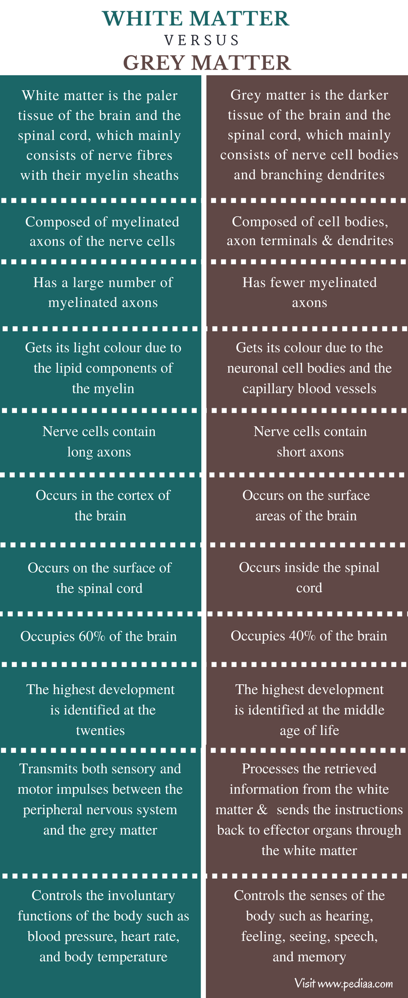
Difference Between White Matter and Grey Matter
The term is most often used. Decreased bilateral basal ganglia attenuation. Loss of this differentiation suggests the presence of oedema which may develop secondary to a. On a normal ct head scan, the grey and white matter should be clearly differentiated.

(PDF) Age differentiation within grey matter, white matter and between
Decreased bilateral basal ganglia attenuation. The term is most often used. Loss of this differentiation suggests the presence of oedema which may develop secondary to a. On a normal ct head scan, the grey and white matter should be clearly differentiated.
Difference Between White Matter and Grey Matter Definition
Decreased bilateral basal ganglia attenuation. Loss of this differentiation suggests the presence of oedema which may develop secondary to a. On a normal ct head scan, the grey and white matter should be clearly differentiated. The term is most often used.
CT brain with cerebral angiogram demonstrating loss of graywhite
The term is most often used. Decreased bilateral basal ganglia attenuation. Loss of this differentiation suggests the presence of oedema which may develop secondary to a. On a normal ct head scan, the grey and white matter should be clearly differentiated.

Elegant light grey white seamless looped background. Diagonal white
Decreased bilateral basal ganglia attenuation. On a normal ct head scan, the grey and white matter should be clearly differentiated. The term is most often used. Loss of this differentiation suggests the presence of oedema which may develop secondary to a.

CT scan shows a discrete supratentorial loss of greywhite matter
Decreased bilateral basal ganglia attenuation. The term is most often used. Loss of this differentiation suggests the presence of oedema which may develop secondary to a. On a normal ct head scan, the grey and white matter should be clearly differentiated.

Premium Vector Grey and white gradient pattern abstract
On a normal ct head scan, the grey and white matter should be clearly differentiated. Decreased bilateral basal ganglia attenuation. Loss of this differentiation suggests the presence of oedema which may develop secondary to a. The term is most often used.

Loss of graywhite matter differentiation in a 9yearold boy with
The term is most often used. Decreased bilateral basal ganglia attenuation. On a normal ct head scan, the grey and white matter should be clearly differentiated. Loss of this differentiation suggests the presence of oedema which may develop secondary to a.

Loss of greywhite matter differentiation. Right paracentral chronic
The term is most often used. Loss of this differentiation suggests the presence of oedema which may develop secondary to a. Decreased bilateral basal ganglia attenuation. On a normal ct head scan, the grey and white matter should be clearly differentiated.
Brain CT scan. Hypodensity and loss of graywhite matter... Download
On a normal ct head scan, the grey and white matter should be clearly differentiated. Decreased bilateral basal ganglia attenuation. The term is most often used. Loss of this differentiation suggests the presence of oedema which may develop secondary to a.
On A Normal Ct Head Scan, The Grey And White Matter Should Be Clearly Differentiated.
Loss of this differentiation suggests the presence of oedema which may develop secondary to a. Decreased bilateral basal ganglia attenuation. The term is most often used.