Poorly Differentiated Squamous Cell - In this paper, we present a challenging and complexly interpreted case of a lesion with some cytohistologic features of. No / minimal keratinization, marked nuclear atypia, may be difficult to establish squamous differentiation. However, 20 to 25 percent of cups are poorly differentiated and cannot be precisely characterized by histologic examination.
However, 20 to 25 percent of cups are poorly differentiated and cannot be precisely characterized by histologic examination. No / minimal keratinization, marked nuclear atypia, may be difficult to establish squamous differentiation. In this paper, we present a challenging and complexly interpreted case of a lesion with some cytohistologic features of.
In this paper, we present a challenging and complexly interpreted case of a lesion with some cytohistologic features of. No / minimal keratinization, marked nuclear atypia, may be difficult to establish squamous differentiation. However, 20 to 25 percent of cups are poorly differentiated and cannot be precisely characterized by histologic examination.

Poorly differentiated squamous cell carcinoma with ulceration
No / minimal keratinization, marked nuclear atypia, may be difficult to establish squamous differentiation. In this paper, we present a challenging and complexly interpreted case of a lesion with some cytohistologic features of. However, 20 to 25 percent of cups are poorly differentiated and cannot be precisely characterized by histologic examination.
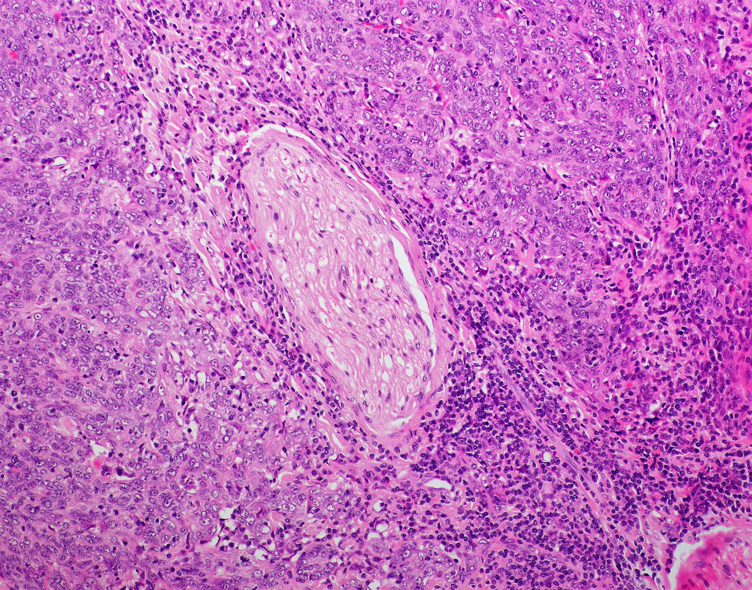
Poorly differentiated squamous cell carcinoma of the larynx with
In this paper, we present a challenging and complexly interpreted case of a lesion with some cytohistologic features of. However, 20 to 25 percent of cups are poorly differentiated and cannot be precisely characterized by histologic examination. No / minimal keratinization, marked nuclear atypia, may be difficult to establish squamous differentiation.
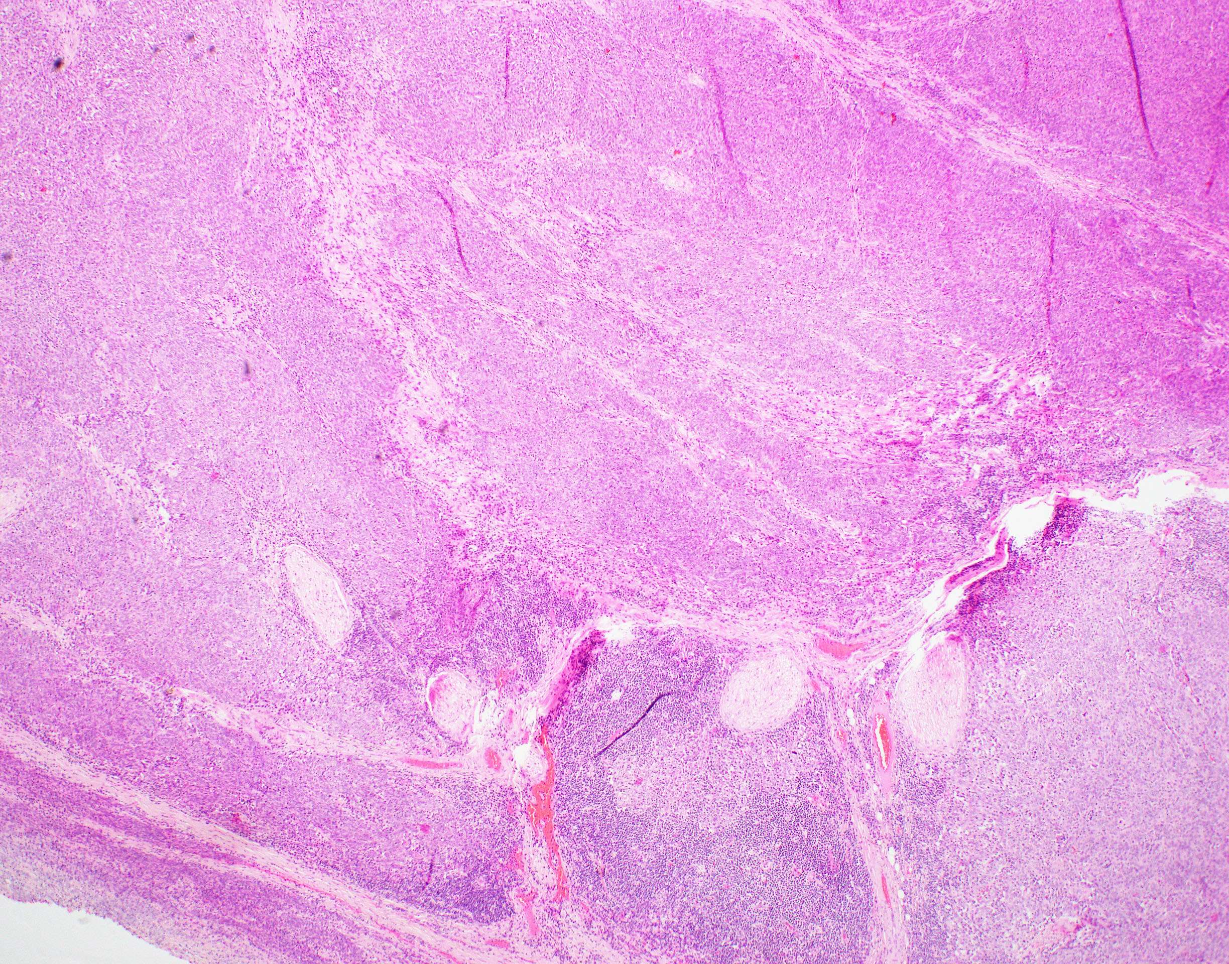
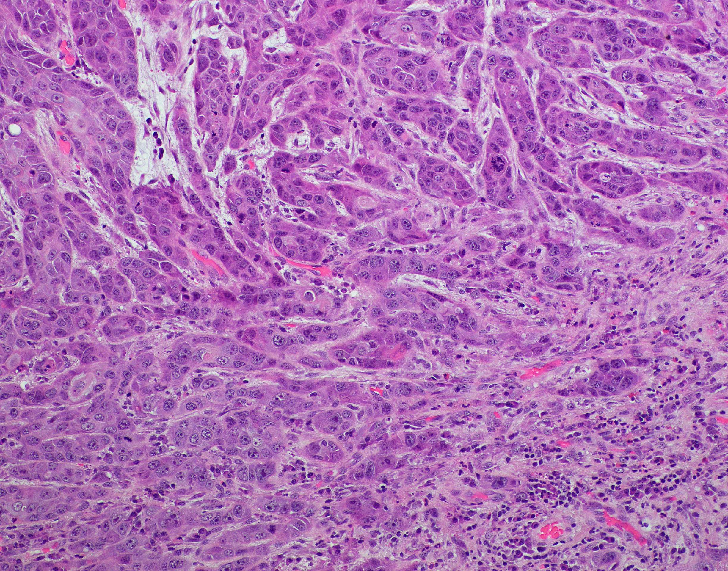
Poorly differentiated squamous cell carcinoma pathology image
In this paper, we present a challenging and complexly interpreted case of a lesion with some cytohistologic features of. No / minimal keratinization, marked nuclear atypia, may be difficult to establish squamous differentiation. However, 20 to 25 percent of cups are poorly differentiated and cannot be precisely characterized by histologic examination.

moderately to poorly differentiated squamous cell cancer. Download
In this paper, we present a challenging and complexly interpreted case of a lesion with some cytohistologic features of. No / minimal keratinization, marked nuclear atypia, may be difficult to establish squamous differentiation. However, 20 to 25 percent of cups are poorly differentiated and cannot be precisely characterized by histologic examination.
Poorly differentiated squamous cell carcinoma of the larynx with
No / minimal keratinization, marked nuclear atypia, may be difficult to establish squamous differentiation. In this paper, we present a challenging and complexly interpreted case of a lesion with some cytohistologic features of. However, 20 to 25 percent of cups are poorly differentiated and cannot be precisely characterized by histologic examination.
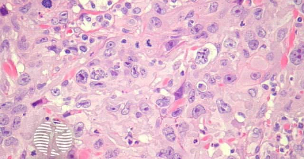
Poorly Differentiated Squamous Carcinoma, Small Cell Variant
However, 20 to 25 percent of cups are poorly differentiated and cannot be precisely characterized by histologic examination. In this paper, we present a challenging and complexly interpreted case of a lesion with some cytohistologic features of. No / minimal keratinization, marked nuclear atypia, may be difficult to establish squamous differentiation.

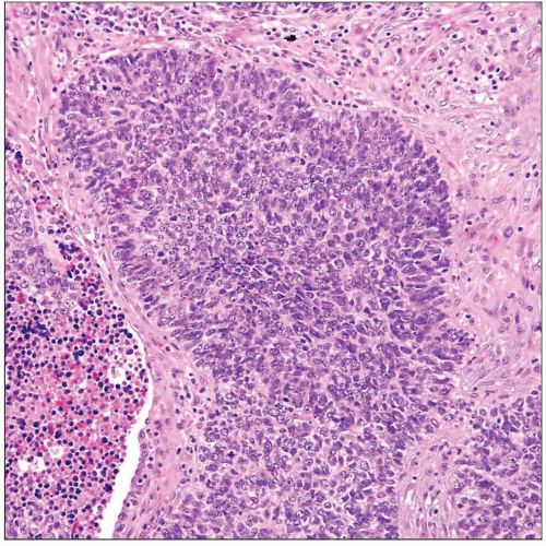
15 Squamous cell carcinoma. Poorly differentiated (nonkeratinizing
No / minimal keratinization, marked nuclear atypia, may be difficult to establish squamous differentiation. In this paper, we present a challenging and complexly interpreted case of a lesion with some cytohistologic features of. However, 20 to 25 percent of cups are poorly differentiated and cannot be precisely characterized by histologic examination.
Moderately differentiated squamous cell carcinoma of the floor of oral
No / minimal keratinization, marked nuclear atypia, may be difficult to establish squamous differentiation. However, 20 to 25 percent of cups are poorly differentiated and cannot be precisely characterized by histologic examination. In this paper, we present a challenging and complexly interpreted case of a lesion with some cytohistologic features of.

Poorly Differentiated Squamous Cell Carcinoma Skin Stock Photo
No / minimal keratinization, marked nuclear atypia, may be difficult to establish squamous differentiation. In this paper, we present a challenging and complexly interpreted case of a lesion with some cytohistologic features of. However, 20 to 25 percent of cups are poorly differentiated and cannot be precisely characterized by histologic examination.

Poorly differentiated squamous cell carcinoma. Smear shows cohesive
In this paper, we present a challenging and complexly interpreted case of a lesion with some cytohistologic features of. No / minimal keratinization, marked nuclear atypia, may be difficult to establish squamous differentiation. However, 20 to 25 percent of cups are poorly differentiated and cannot be precisely characterized by histologic examination.
No / Minimal Keratinization, Marked Nuclear Atypia, May Be Difficult To Establish Squamous Differentiation.
In this paper, we present a challenging and complexly interpreted case of a lesion with some cytohistologic features of. However, 20 to 25 percent of cups are poorly differentiated and cannot be precisely characterized by histologic examination.